 Much can be written about Value-based care. I’ll focus on imaging risk management from a radiologist’s perspective. What it looks like from the Hospital’s perspective , the Insurer’s perspective, and in general have been discussed previously.
When technology was in shorter supply, radiologists were gatekeepers of limited Ultrasound, CT and MRI resources. Need-based radiologist approval was necessary for ‘advanced imaging’. The exams were expensive and needed to be protocoled correctly to maximize utility. This encouraged clinician-radiologist interaction – thus our reputation as “The Doctor’s doctor.”
In the 1990’s-2000’s , there was an explosion in imaging utilization and installed equipment. Imaging was used to maximize throughput, minimize patient wait times and decrease length of hospital stays. A more laissez-faire attitude prevailed where gatekeeping was frowned upon.
With a transition to value-based care, the gatekeeping role of radiology will return. Instead of assigning access to imaging resources on basis of limited availability, we need to consider ROI (return on investment) in the context of whether the imaging study will be likely to improve outcome vs. cost. (1) Clinical Decision Support (CDS) tools can help automating imaging appropriateness and value. (2)
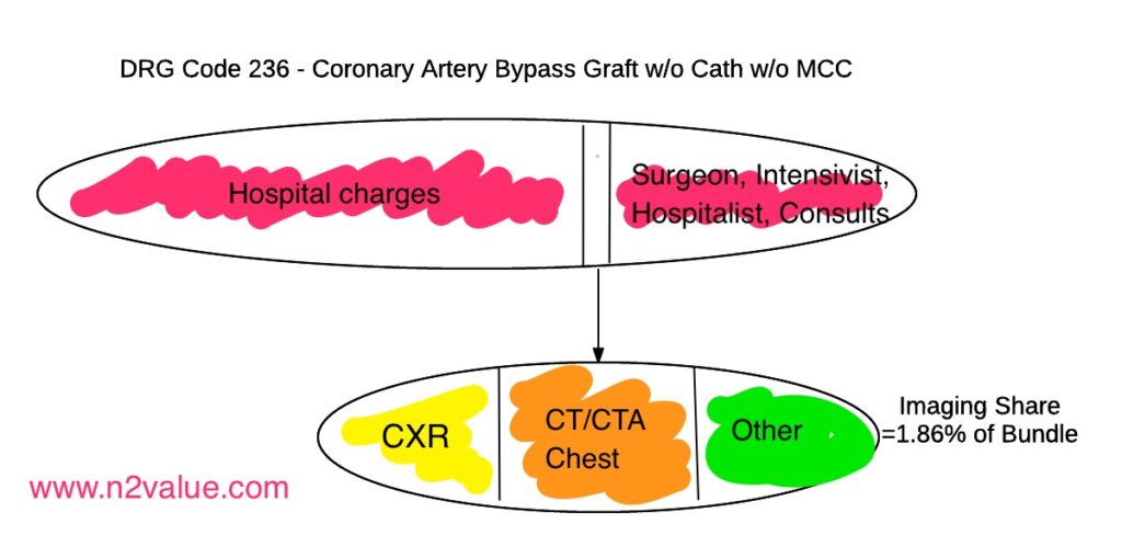
The bundle’s economics are capitation of a single care episode for a designated ICD-10 encounter. This extends across the inpatient stay and related readmissions up to 30 days after discharge (CMS BPCI Model 4). A review of current Model 4 conditions show mostly joint replacements, spinal fusion, & our example case of CABG (Coronary Artery Bypass Graft).
Post CABG, a daily Chest X-ray (CXR) protocol may be ordered – very reasonable for an intubated & sedated patient. However, an improving non-intubated awake patient may not need a daily CXR. Six Sigma analysis would empirically classify this as waste – and a data analysis of outcomes may confirm it.
Imaging-wise, patients need a CXR preoperatively, & periodically thereafter. A certain percentage of patients will develop complications that require at least one CT scan of the chest. Readmissions will also require re-imaging, usually CT. There will also be additional imaging due to complications or even incidental findings if not contractually excluded (CT/CTA/MRI Brain, CT/CTA neck, CT/CTA/US/MRI abdomen, Thoracic/Lumbar Spine CT/MRI, fluoroscopy for diaphragmatic paralysis or feeding tube placement, etc…). All these need to be accounted for.
In the fee-for-service world, the ordered study is performed and billed. In bundled care, payments for the episode of care are distributed to stakeholders according to a pre-defined allocation.
Practically, one needs to retrospectively evaluate over a multi-year period how many and what type of imaging studies were performed in patients with the bundled procedure code. (3) It is helpful to get sufficient statistical power for the analysis and note trends in both number of studies and reimbursement. Breaking down the total spend into professional and technical components is also useful to understand all stakeholder’s viewpoints. Evaluate both the number of studies performed and the charges, which translates into dollars by multiplying by your practice’s reimbursement percentage. Forward-thinking members of the Radiology community at Nieman HPI are providing DRG-related tools such as ICE-T to help estimate these costs (used in above image). Ultimately one ends up with a formula similar to this:
CABG imaging spend = CXR’s+CT Chest+ CTA chest+ other imaging studies.
Where money will be lost is at the margins – patients who need multiple imaging studies, either due to complications or incidental findings. With between a 2% to 3% death rate for CABG and recognizing 30% of all Medicare expenditures are caused by the 5% of beneficiaries that die, with 1/3 of that cost in the last month of life (Barnato et al), this must be accounted for. An overly simplistic evaluation of the imaging needs of CABG will result in underallocation of funds for the radiologist, resulting in per-study payment dropping – the old trap of running faster to stay in place.
Payment to the radiologist could either be one of two models:
First, fixed payment per RVU. Advantageous to the radiologist, it insulates from risk-sharing. Ordered studies are read for a negotiated rate. The hospital bears the cost of excess imaging. For a radiologist in an independent private practice providing services through an exclusive contract, allowing the hospital to assume the risk on the bundle may be best.
Second, a fixed (capitated) payment per bundled patient for imaging services may be made to the radiologist. This can either be in the form of a fixed dollar amount or a fixed percentage of the bundle. (Frameworks for Radiology Practice Participation, Nieman HPI) This puts the radiologist at-risk, in a potentially harmful way. The disconnect is that the supervising physicians (cardio-thoracic surgeon, intensivist, hospitalist) will be focusing on improving outcome, decreasing length of stay, or reducing readmission rates, not imaging volume. Ordering imaging studies (particularly advanced imaging) may help with diagnostic certitude and fulfill their goals. This has the unpleasant consequence of the radiologist’s per study income decreasing when they have no control over the ordering of the studies and, in fact, it may benefit other parties to overuse imaging to meet other quality metrics. The radiology practice manager should proceed with caution if his radiologists are in an employed model but the CT surgeon & intensivists are not. Building in periodic reviews of expected vs. actual imaging use with potential re-allocations of the bundle’s payment might help to curb over-ordering. Interestingly, in this model the radiologist profits by doing less!
Where the radiologist can add value is in analysis, deferring imaging unlikely to impact care. Reviewing data and creating predictive analytics designed to predict outcomes adds value while, if correctly designed, avoiding more than the standard baseline of risk. (see John’s Hopkins Sepsis prediction model). In patients unlikely to have poor outcomes, additional imaging requests can be gently denied and clinicians reassured. I.e. “This patient has a 98% chance of being discharged without readmission. Why a lumbar spine MRI?” (c.f. AK Moriarty et al) Or, “In this model patients with these parameters only need a CXR every third day. Let’s implement this protocol.” The radiologist returns to a gatekeeping role, creating value by managing risk, intelligently.
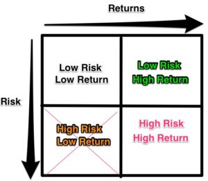
Let’s return to our risk/reward matrix:
For the radiologist in the bundled example receiving fixed payments:
Low Risk/Low Reward: Daily CXR’s for the bundled patients.
High Risk/Low Reward: Excess advanced imaging (more work for no change in pay)
High Risk/High Reward: Arbitrarily denying advanced imaging without a data-driven model (bad outcomes = loss of job, lawsuit risk)
Low Risk/High Reward: Analysis & Predictive modeling to protocol what studies can be omitted in which patients without compromising care.
I, and others, believe that bundled payments have been put in place not only to decrease healthcare costs, but to facilitate transitioning from the old FFS system to the value-based ‘at risk’ payment system, and ultimately capitated care. (Rand Corp, Technical Report TR-562/20) By developing analytics capabilities, radiology providers will be able to adapt to these new ‘at-risk’ payment models and drive adjustments to care delivery to improve or maintain the community standard of care at the same or lower cost.
- B Ingraham, K Miller et al. Am Coll Radiol 2016 in press
- AK Moriarty, C Klochko et al J Am Coll Radiol 2015;12:358-363
- D Seidenwurm FJ Lexa J Am Coll Radiol 2016 in press

